DIAPHRAGM EVENTRATION
Professor İrfan
Yalçınkaya, MD
Health Sciences
University, Istanbul Hamidiye Faculty of Medicine, Department of Chest Surgery,
Sureyyapasa Chest Diseases and Chest Surgery Health and Application Research
Center, Chest Surgery Clinic
E-mail: profdrirfanyalcinkaya@gmail.com
Summary:
In symptomatic patients with diaphragmatic eventration, minimally invasive
plication is the primary option.
Keywords:
Diaphragmatic Eventration, Plication, Video-Assisted Thoracoscopic Surgery,
Minimally Invasive Surgery
Definition
& History & Etiology
Diaphragmatic
eventration is the permanent elevation of all or part of the diaphragm leaf,
provided that the costal parts and organ connections are not disrupted. Another
definition is the permanent elevation of the hemidiaphragm without any defect
in the muscle attachments of the diaphragm and without disruption of the
integrity between the pleural and peritoneal cavities. The terms eventration
and paralysis are often confused. Paralysis describes the muscular degeneration
resulting from denervation and is one of the pathologies that initiate
eventration. However, eventration does not always occur together with
paralysis. Despite this, eventration is used simultaneously to describe both
paralysis and elevation of the diaphragm.
Diaphragm
height was first defined by Jean Louis Petit and published in 1760. The term
“diaphragm eventration” was first used by Beclard in 1829.
While it is not possible
to reveal the etiological cause in the majority of patients (idiopathic
diaphragmatic eventration), malignancy, trauma, infections, iatrogenic causes
(mediastinal and cardiac surgery) and neuromuscular diseases are important etiological
causes. Although the cause cannot always be fully explained, congenital and
acquired diaphragmatic eventration are divided into two groups: those related
to phrenic nerve paralysis and those not. It is one of the rare pathologies
that is often detected incidentally after trauma or during the investigation of
dyspnea etiology.1 Although the cause can be revealed in many
patients, the cause cannot be fully revealed in some patients. The underlying
cause in these patients is thought to be viral infections.2
The main cause of congenital diaphragm eventration is a
congenital developmental defect of the diaphragm muscle, characterized by a
decrease in the amount of muscle fibers. Acquired diaphragm elevation can be
divided into two categories: paralytic and non-paralytic. The main cause of
paralytic diaphragm eventration is the failure of the phrenic nerve to function
after trauma, tumor, or surgery. In cases of phrenic nerve damage, diaphragm
elevation is observed on the affected side (Table 1).
Table
1. Other factors causing diaphragm elevation other than eventration

Diagnosis
Diaphragmatic
eventration, although rare, is more common in men and is usually predominantly
left-sided. The most common symptom of acquired diaphragmatic eventration in
adult patients is respiratory problems. Since the diaphragm is the primary
respiratory muscle, the majority of patients complain of increased dyspnea,
especially with exertion and when lying supine. Less frequently, cough,
retrosternal or epigastric pain may also be a symptom. Respiratory
insufficiency symptoms may also occur due to underlying lung disease or
decreased lung reserve. In addition, nausea, vomiting, abdominal gas-bloating,
belching or abnormally increased bowel sounds may be present as symptoms. It
may also be associated with sleep disorders. Therefore, polysomnography should
be performed in patients with diaphragmatic eventration/paralysis who show
symptoms and who also have sleep disorders. Patients diagnosed with obstructive
sleep apnea syndrome may benefit from surgery, and symptoms may completely
improve or be alleviated. In this way, long-term comorbidities of sleep disorders
can also be prevented.3,4
Diagnosis
is often made incidentally, most of the time, with posteroanterior (PA) or
lateral chest radiography. While an elevation of the diaphragm on plain
radiography may not indicate diaphragmatic paralysis, paralysis can be excluded
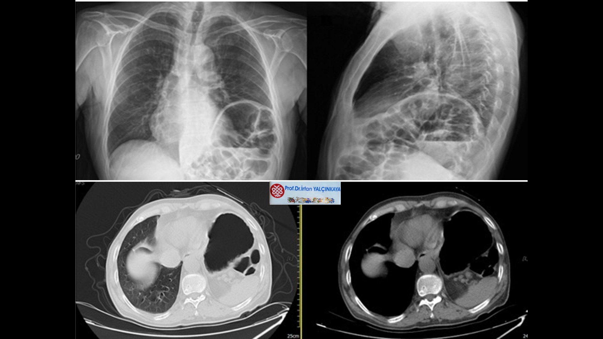
if an elevated diaphragm is not seen. Thoracic Computed Tomography (CT) and,
when necessary, Ultrasonography are also valuable methods in demonstrating the
underlying pathology (Figure 1). However, these methods have limited ability to
distinguish diaphragmatic eventration and hernias. Magnetic Resonance Imaging
can also be used when necessary.
Figure
1: Left diaphragm eventration (PA chest X-ray, Left lateral chest X-ray), left
diaphragm eventration in thorax CT

If
the phrenic nerve is affected, diaphragm movements are reduced or absent on
fluoroscopy, or paradoxical movements of the diaphragm can be seen. In patients
who are suddenly inspired during fluoroscopy, the normal diaphragm moves
downwards, while the paralyzed diaphragm moves upwards. Fluoroscopy or
ultrasonography should definitely be performed, especially in cases with right
diaphragm eventration. Today, fluoroscopy has almost completely given way to
ultrasonography. Diaphragm thickness can be easily measured and diaphragm
movements can be evaluated with thoracic ultrasonography. It should not be
forgotten that pathologies under the diaphragm (such as hepatomegaly,
subphrenic abscess, intra-abdominal pathologies) can cause right diaphragm
elevation, and pleural effusion and pericardial cysts can also mimic diaphragm
eventration.
Pulmonary
Function Tests (PFTs) can be helpful in the evaluation of dyspneic patients with
diaphragmatic eventration, and these patients often have decreased FEV1 and FVC
values. Since the dysfunctional diaphragm cannot retract sufficiently, there is
a decrease in lung volumes and compliance. This is reflected in spirometric
values, and FVC and FEV1 values are decreased. Although PFT values are
usually abnormal in symptomatic patients with diaphragmatic
eventration/paralysis, these values are often not related to the severity of
dyspnea. PFTs are the most objective criteria used to exclude other factors in
the differential diagnosis of dyspnea, in preoperative evaluation in
eventration, in postoperative follow-up, and in response to surgery.
In
the differential diagnosis, the causes of exertional dyspnea and increase in
dyspnea, such as Chronic Obstructive Pulmonary Disease, congestive heart
failure, and morbid obesity, should be carefully examined.
Treatment
In
the presence of symptomatic diaphragmatic eventration & paralysis,
treatment is surgical. Regardless of etiology, plication is the basic surgical
method.2,5
The
aim of surgical plication is to stabilize the atrophic, thin, flaccid and
elevated diaphragm, preventing the abdominal organs from moving to the
evanescent side during inspiration, and to prevent the development of parenchymal
linear atelectasis due to the compression of the mediastinal shift and elevated
diaphragm. In addition, the elevation in the diaphragm is reduced to the normal
position by plication, and the intrathoracic negative pressure required for the
expansion of the contralateral lung is re-regulated, thus improving exercise
performance.2,6
In
asymptomatic cases and secondary eventrations caused by neoplasm, even if
eventration is detected on chest X-ray, there is no indication for surgery
unless it is symptomatic and necessary. (Figure 2)
Figure
2. A case without surgical indication

Surgery
is indicated when respiratory symptoms (usually dyspnea, especially dyspnea on
exertion or orthopnea, less frequently cough) or when gastrointestinal findings
(dyspepsia, meteorism) are at a level that will affect the patient's life.
After neoplasia is excluded, surgery can be decided by considering the
patient's clinical severity and duration of symptoms. If the cause of
eventration is paralysis and the symptoms are new, it can be waited for 6-12
months for the picture to become clear in these patients, and in cases other
than paralysis, we believe that there is no need to wait for surgery in
patients with clinical symptoms (dyspnea on exertion) and radiologically
demonstrated eventration. Spontaneous resolution is possible, albeit rarely, in
eventrations that occur in the early postoperative period. Asymptomatic cases
should be monitored and surgical treatment should be considered in case of
symptoms or deterioration of respiratory functions. In addition, when phrenic
nerve injury develops in patients undergoing thoracic surgery (especially in
pneumonectomy), prophylactic diaphragm plication can be performed at the same
session to reduce future respiratory complications.7 For patients on
ventilator support, plication is not contraindicated if indicated.
Wood
first proposed the idea that the diaphragm could be reduced in size by
wrinkling it in 1916. In 1923, Morrison performed the first successful
eventration surgery and defined the surgical principle that is still used.
Many
methods have been defined in diaphragmatic eventration surgery and have been
used throughout history. The traditional method for patients with symptomatic
diaphragmatic eventration is open transthoracic or transabdominal plication.
This method can provide significant improvement in patients' symptoms and
considerable improvement in respiratory functions. However, open surgery is
invasive and can also cause certain morbidity and even mortality. It is not a
good option, especially in patients with multiple comorbidities and poor lung
capacity. In addition, no matter how small the incision used in this surgery,
which is performed to reduce and eliminate respiratory distress, it can have a
restrictive effect on both pain and respiratory functions in the postoperative
period, thus reducing the success of the expected outcome of the surgery.2
In
order to minimize the disadvantages of open transthoracic surgery, Gharagozloo
et al. performed the first thoracoscopic diaphragm plication in a 72-year-old
patient with left phrenic nerve involvement and left diaphragm paralysis due to
thymic carcinoma in 1995. This procedure is technically no different from the
classical transthoracic approach, except that it is performed with video
assistance. In 1996, Mouroux et al. developed and published a thoracoscopic
method that is technically more applicable than Gharagozloo's method.8
The
first VATS applications were mostly in infants and children.9 In the
literature, in the first series of publications on adults, cases where
thoracotomy and VATS were applied are mostly together. Today, open surgical
methods have given way to minimally invasive surgical methods. Open surgery is
preferred only in patients where intubation with a double-lumen tube cannot be
applied for VATS, or the patient cannot tolerate it, or in patients who cannot
perform minimally invasive surgery due to advanced pleural/peritoneal adhesions
due to previous diseases and surgeries. (Table 2)
Table
2. Surgical methods in diaphragm pathologies

In
the VATS approach, the patient is positioned in the lateral decubitus position
and 30 degrees reverse Trendelenburg, as in open surgery. The first port is
opened from the 6th intercostal space (ICA) in the midaxillary line and the
location of the other 2 ports is determined after diaphragm exploration, and
some surgeons use CO2 to depress the diaphragm. During surgery, plication is
performed using only thoracoscopic instruments (Endograsper, Endostitch, etc.).
In the literature, the number of ports used for VATS varies between 2 and 4,
and it is stated that this procedure can be performed with a single port.10
Although the endoscopic instruments used in the VATS procedure are suitable for
diaphragm eventration in infants and children, it is relatively more difficult
to perform a plication with sufficient tension with endoscopic instruments in
adults because the diaphragm has both a larger surface area and a thicker
thickness.
In
diaphragm plication with RATS, the patient is in the lateral decubitus
position, the head is in a 10-degree reverse Trendelenburg position. The first
port is made from the place where the diaphragm is thought to be the highest,
and CO2 is injected into the intrathoracic cavity. Then, the camera port is
opened from the 4th ICA and after exploration, 2 more ports are opened for the
robot arms and plication is applied. Although 3-dimensional image and no
restriction of movement during plication seem to be among the advantages, the
high cost of RATS, its not being easily accessible and widespread can be
considered among the disadvantages of this method.
Another
method is the hybrid method and is called video-assisted mini-thoracotomy. This
method is a plication performed with 1 camera port and 1 utility thoracotomy.
Thus, open surgical instruments can be used easily and the procedure can be
performed with the comfort of open surgery. The intrathoracic area can be
illuminated and when the diaphragm is grasped and pulled, the structures under
the diaphragm can be easily seen with transillumination. It is not easy to
suture the diaphragm with sufficient thickness and length with endostitch,
which makes it difficult to create sufficient tension. Moreover, the 3-4 port
sites used in VATS can reach a total utility incision. This method, which was
first described by Lai and Paterson in 1999 and reported as a single case, was
later modified by Rombolá et al. and applied in 18 cases.10,11
We
have successfully applied a modified version of these two methods with
different characteristics in cases with diaphragmatic eventration.12-16
In
this method, the patient was intubated under general anesthesia with a
double-lumen intubation tube according to the side. The patients were laid on
the operating table in the lateral decubitus position with the operation side
uppermost, with the head 30-45 degrees higher. In this way, the abdominal
organs were taken down and the pressure of the abdominal organs on the
diaphragm was reduced. The arm on the operating side was positioned in a way
that would not hinder the movement of the endocamera. A nasogastric tube was
inserted to provide stomach decompression and reduce abdominal distension. This
procedure is especially important on the left side, thus allowing the diaphragm
to descend more easily and reducing tension, allowing easier maneuverability
for the surgeon. It may not be performed on the right side, but it can be. In
these patients, an additional bowel cleansing is not necessary.
The
surgeon who will perform the operation takes his/her place behind the patient
if the eventration is on the right and in front of him/her if the eventration
is on the left. First, a 10 mm (5 mm) port hole was opened for the endocamera
from the 3rd or 4th ICA in the axillary line. The thoracic cavity and the
eventrated diaphragm were evaluated using a zero-degree optic (30 degree).
Unilateral ventilation was initiated and the lung on the side where the
plication would be applied was deflated. The location where the thinned and
eventrated diaphragm adhered to the chest wall was determined and a 3-4 cm
utility thoracotomy was opened from the mid-axillary line two ribs above
(usually the 7th or 8th ICA). In this way, both the level at which the
diaphragm could be tense was determined (for this, a transthoracic syringe
needle tip can be used if necessary) and a suitable and sufficient area was
provided for working with the instruments used in open surgery (or preferably
open instruments developed for thoracoscopy). In utility thoracotomy
retraction, initially pediatric retractors and Weitlaner retractors were used,
and later wound protection retractors (Alexis etc.) were used in all cases.
The
stitches were first placed with a needle and forceps, starting from the closest
point where the diaphragm adheres to the chest wall, from lateral to medial,
and starting from the point where the pericardium sits on the diaphragm, using
a needled number 1 silk suture in a continuous single row. The first sutures
can be placed directly by seeing the diaphragm directly from the retractor and
applying a sling with the help of the sutures, while the final sutures can be
placed easily by looking at the monitor. The diaphragm was lifted with forceps
or clamps and plication was applied without damaging the abdominal organs with
the help of the camera light and the transillumination method. When the
plication process was most medial, the plication line was tightened like a
spring from both suture ends with the help of a finger and a knot was tied at the
closest point to the utility thoracotomy, and a knot pusher was used when
necessary. With this first row of sutures, other plication lines became
apparent, and subsequent procedures became easier. Then, in the same way, two
rows of stitches were sewn on both sides of this first row of stitches to
provide sufficient tension in the diaphragm, and the diaphragm was lowered to
its normal position.
If
the patient's diaphragm is extremely thin and it is thought that the sutures
may tear, a pledget suture may be used. During plication, care was taken not to
try to pass the needle through the folds formed when the diaphragm, which is
loose and loose, was held with forceps and not to insert the needle either
superficially or too deeply. In this way, the diaphragm was plicated very
strongly and a possible complication related to the structures under the
diaphragm was prevented. After checking the diaphragm tension with a finger or
a tool, the entire field was reviewed with the help of a camera. Care should be
taken not to over-tension the diaphragm as a result of plication. Because the
sutures placed may tear in cases where intra-abdominal pressure increases.
A
chest tube of size 24 or 28 was inserted into the pleural space through a
utility thoracotomy and placed in the pleural cavity with its tip at the apex.
The incision should be closed very well, otherwise lung herniation may occur,
and I believe that a single intercostal suture may be placed if deemed
necessary. Postoperative PA chest radiographs were taken for all patients to
see the diaphragm levels in the early period. Nasogastric tube was terminated
with the onset of bowel movements on the first postoperative day and all
patients were started on a soft diet. If there is no drainage & air leak
and the lung is expanded on the PA chest radiograph, the drain can usually be
removed at the end of the first or second postoperative day. The patient whose
drain has been removed can be discharged after a follow-up chest radiograph.
After discharge, the patient is advised not to be constipated for a while and
not to lift heavy objects.
Many
methods are described in the literature for diaphragm plication; hand-tied U
sutures, matrix sutures, continuous sutures with or without pledgets or
staples, methods with or without mesh, resection of a portion of the diaphragm
and bringing the remaining portion end to end with overlapping sutures.8,14
Although
there is no study comparing VATS and plication methods, a study by Evman et al.
compared plication techniques performed via thoracotomy. In a series of 42
cases, accordion plication and laparoscopic plication techniques were compared
and increases in spirometric values were found to be similar between the two
groups. It was emphasized that there was an increase in spirometric tests
regardless of the technique.17 The choice of plication method
depends on the surgeon's expertise, training, and preference. Improvement in
dyspnea is the most important measure of clinical success.
We
apply and prefer accordion plication with non-absorbable continuous interrupted
sutures such as silk. However, prolene and polyester sutures can also be used
as suture materials. We also used pledget sutures in a few cases where the
diaphragm was extremely thinned and we thought it would rupture with the
sutures. We do not prefer staples due to concerns that the staples may break as
a result of the tension created in the diaphragm, and we also do not prefer
diaphragm resection due to the need for sufficiently thick tissue, even if it
is thinned. We do not recommend mesh and think that it can only be used in
recurrent cases.
In
cases of right-sided diaphragmatic eventration, one must be very careful and
meticulous. The cause of eventration must be investigated thoroughly, and if
possible, fluoroscopy must be performed. Since fluoroscopy is no longer widely
used, in recent years we have preferred diaphragmatic ultrasonography. Thus,
the movement and thickness of the diaphragm can be easily measured. The
patient's symptoms and respiratory function values should be carefully
examined in terms of COPD, obesity and heart failure. While no indication for
surgery was given in four cases with right diaphragmatic eventration, in two
cases the diaphragm was found to be normal during surgery and plication was
abandoned.
Even
in cases with a history of trauma, when thoracotomy is performed, it is
possible to encounter eventration instead of rupture in the diaphragm. In fact,
different pathologies such as diaphragmatic hernia and pericardial cyst can be
encountered. Therefore, even if there is a history of trauma, we recommend
starting all diaphragmatic pathologies with VATS. Because in closed surgery, if
desired and necessary, it is possible to convert to open surgery at any time,
but in open surgery, it is not possible to convert to closed surgery.
In
the Chest Surgery Clinic of the Health Sciences University, Istanbul
Sureyyapasa Chest Diseases and Chest Surgery Health Application and Research
Center, between December 2009 and May 2025, 106 cases with symptoms of exertional
dyspnea and diagnosed with diaphragm paralysis & eventration underwent
minimally invasive surgery with diaphragm plication. In all cases, clinical
complaints improved completely or largely in the postoperative period. No
mortality was observed, and more than ten cases developed minimal complications
(lung herniation in one case), and recurrence was seen in one case, and
follow-up was sufficient. (Figure 3)
Figure
3. Our 106th case with VATS plication

In
the doctoral thesis study of 50 cases titled “The effect of minimally invasive
surgery on respiratory functions in diaphragmatic eventration” conducted in
2017 by Deniz Gürer MD and for which I was the thesis advisor at the same
institution; when the preoperative and postoperative FEV1 (% and lt) values
of the patients were compared, a statistically significant increase was found
in both parameters (p<0.001). There was an average increase of 12.1% in FEV1
(lt) values. Similarly, when the preoperative and postoperative FVC (% and lt)
values were compared, a statistically significant increase was found in both
parameters (p<0.001). There was a postoperative increase of 11.5% in FVC
(lt) values. Recurrence was detected in one patient during long-term
follow-ups; this patient did not accept surgery and was followed up.
Surgery
is indicated in symptomatic cases with diaphragmatic eventration and effort
dyspnea, especially on the left. Today, diaphragmatic plication with minimally
invasive surgery is an easy, fast and highly effective method in diaphragmatic
pathologies, especially in diaphragmatic eventration, as in other thoracic
structures. In this surgical method, where the instruments used in open surgery
can be used, sufficient diaphragmatic tension similar to that in open surgery
can be achieved and statistically significant improvements can be achieved in
spirometric tests. At the same time, as in other surgeries performed with
minimally invasive surgery, postoperative pain and morbidity are less, patient
comfort increases, the recovery process is accelerated and the patient is
discharged quickly.18-21
We
would like to underline once again that plication with minimally invasive
surgery is the primary option in symptomatic patients with diaphragmatic
eventration.
*Viewing
Recommendation: Minimally Invasive Surgery in Diaphragmatic Eventration, https://www.youtube.com/watch?v=GKqM4OX37Xs
*Viewing
Recommendation: Prof. Dr. İrfan Yalçınkaya, Minimally Invasive Surgery in
Diaphragmatic Eventration & Paralysis, School of Thoracic Surgery, 2024,
https://www.youtube.com/watch?v=3DOxq0VrRRU&t=245s
References:
1.
Yalçınkaya İ. Dispne etyolojisinin araştırılması sırasında saptanan nadir bir
patoloji: Diyafram evantrasyonu [A rare pathology detected during the
investigation of dyspnea etiology: Diaphragm eventration]. Akciğer Bülteni
2019;5(2):51-5.
2.
Yalçınkaya İ. Diyafram paralizisi ve evantrasyonu [Diaphragm
paralysis and eventration]. Yalçınkaya İ, editör. Diyafram Hastalıkları. Toraks
Cerrahisi Bülteni 2013;4(4):266-71.
3.
Oruc O, Sarac S, Afsar GC, Topcuoglu OB, Kanbur S, Yalcinkaya I,
Tepetam FM, Kirbas G. Is polysomnographic examination necessary for
subjects with diaphragm pathologies? Clinics
2016;71(9):506-10.
4.
Sarac S, Salturk C, Oruc O, Kanbur Metin S, Bayram S, Karakurt
Z, Yalcınkaya I. Sleep-related breathing disorders in diaphragmatic
pathologies. Sleep and Breathing 2021;26:959-963.
5.
Derdiyok O, Yalçınkaya İ. Diyafram patolojilerinde cerrahi
yaklaşım [Surgical approach to diaphragm pathologies]. In: Göğüs Cerrahisi.
Metin M, Cansever L, Sezen CB (eds). Akademisyen Yayınevi, Ankara, 2022,
Sh.415-20.
6.
Işık AF, Yalçınkaya İ, Kurnaz M. Erişkinlerde diyafragma yükselmeleri;
Cerrahi gerekli mi? [Diaphragmatic elevations in adults; Is surgery necessary?]
Van Medical Journal 2002;9(1):33–7.
7.
Yalçınkaya İ, Doğruyol MT. Prophylactic chest surgery procedures.
In: Dilek ON, Uranues S, Latifi R, eds.
Prophylactic Surgery. 1 st ed. Switzerland: Springer; 2021. p. 371-8.
8.
Gürer D, Atinkaya C, Yalçınkaya İ. Diyafram eventrasyonu [Diaphragm
eventration]. In: Eren TŞ, editor. Torasik Cerrahi. 1st ed. İstanbul: İstanbul
Tıp Kitapevi 2019; p. 53–60.
9.
İrdem AK, Doyurgan O, Kılıç Y, Balık H. İnfantlarda
konjenital kalp cerrahisi sonrasında gelişen diyafram felcinin cerrahi tedavisi
[Surgical treatment of diaphragmatic paralysis developing after congenital
heart surgery in infants]. Dicle
Tıp Dergisi 2024;51(3):415-24.
10. Rombolá C, Crespo
MG, López PT, Martínez AH, Atance PL, Ramírez AT, et al. Video-assisted
minithoracotomy diaphragmatic plication: respiratory effects in adults. Thorac
Cardiovasc Surg. 2016;64(08):647–53.
11. Lai DTM, Paterson
HS. Mini-thoracotomy for diaphragmatic plication with thoracoscopic assistance.
Ann Thorac Surg. 1999;68(6):2364–5.
12. Yalçınkaya İ.
Plikasyon [Plication]. In: Toker A, Batırel HF, editors. Çağdaş
Videotorakoskopik Cerrahide Teknikler ve Sonuçlar. 1st ed. İstanbul: Nobel Tıp
Kitabevi; 2014; p. 157–62.
13. Yalcinkaya I, Evman
S, Lacin T, Alpay L, Kupeli M, Ocakcioglu I. Video-assisted minimally invasive
diaphragmatic plication: feasibility of a recognized procedure through an
uncharacteristic hybrid approach. Surg Endosc. 2017;31(4):1772–7.
14. Yalçınkaya İ,
Atinkaya Baytemir C. Diyafram Eventrasyonu ve Paralizisi ]Diaphragm Eventration
and Paralysis]. In: Yalçınkaya İ, editor. Diyafram Hastalıkları. 1st ed.
Ankara: Türkiye Klinikleri Yayınevi, 2020; p. 38–44.
15. Doğruyol MT, Yalçınkaya
İ. Chapter 75: VATS plication of diaphragm. In: Advanced Thoracic Surgery;
Balcı AE, Ersöz H, Yüksel M (editors), Ankara; Akademisyen Kitabevi,
2021;781-5.
16. Yalçınkaya İ. VATS İle diyafram cerrahisi [Diaphragm
surgery with VATS]. In: Minimal İnvazif Torasik Yaklaşımlar; Metin M, Sezen CB
(editors), İstanbul; Medical Network, 2021;291-308.
17. Evman S, Tezel C, Vayvada M, Kanbur S, Urek S, Baysungur V,
Yalçınkaya I. Comparison of mid-term clinical outcomes of different surgical
approaches in symptomatic diaphragmatic eventration. Ann Thorac Cardiovasc
Surg. 2016;224-29.
18. Nardini M, Jayakumar S, Migliore M, Nosotti M,
Paul I, Dunning J. Minimally invasive plication of the diaphragm: A
single-center prospective study. Innovations 2021;16(4):343-9.
19. Gritsiuta AI, Gordon M, Bakhos CT, Abbas AE,
Petrov RV. Minimally invasive diaphragm plication for acquired unilateral
diaphragm paralysis: A systematic review. Innovations 2022;17(3):180-90.
20. Gilbert A, Wei B. Diaphragmatic plication:
current evidence and techniques in the management of the elevated
hemidiaphragm. Video-Assisted
Thoracic Surgery 2023;8.
21. Balamurugan G, Bhandarwar A, Wagh A, Bakhshi G,
Ansari K, Bhondve S, Dhimole N, Jawale H. Comparison of short-term outcomes of
video-assisted thoracoscopic (VATS) plication of diaphragmatic eventration-a
six-year prospective cohort study. Updates
in Surgery 2024;76(1):279-88.